
MRI* with a Twist Could Show what Life and Disease Really Look Like
Professor Jan Henrik Ardenkjaer-Larsen[1], principal scientist at GE Healthcare, with an honorary professorship at the Technical University of Denmark[2] (DTU), has worked with magnetic resonance (MR) technology for nearly all of his working life. Now, he and his team have been awarded 55 million Danish Kroner by the Danish National Research Foundation[3] to develop his pioneering work. He is using MRI to obtain images of the biochemical reactions of life going on inside us, as they happen, in real time. In the space of ten years, professor Ardenkjaer and his team have taken their technique “from a crazy idea, all the way into man.”
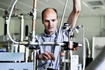
Professor Jan Henrik Ardenkjaer-Larsen, principal scientist at GE Healthcare with an honorary professorship at the Technical University of Denmark (DTU).
Medical imaging is a cornerstone of modern medicine. Without it, doctors would not be able to diagnose disease with the accuracy and precision that X-ray, CT, MRI** and other imaging technologies allow.
MRI, or magnetic resonance imaging, has become especially ubiquitous since its invention over forty years ago. “MRI is a very successful technique, and very widespread,” says Prof Ardenkjaer. It is a very important diagnostic visualization tool for the medical doctor. We can acquire beautiful images in terms of resolution and contrast.”
In the last few years, MRI technology[4] has evolved to the point where we can now create a photo-accurate, three-dimensional computer model[5] of a person’s body, then upload it to the cloud to be examined by physicians virtually anywhere on the planet.
But where MRI is challenged, and where Prof Ardenkjaer et al have been breaking ground for the last decade, is at the fundamental level where all diseases operate: metabolism. “It’s hard to even think of a disease which doesn’t have a metabolic component,” says Professor Ardenkjaer. “Pretty much all disease changes metabolism.”
MRI can provide spectacular images, still and moving, of the gross anatomy of the human body. But the microscopic, biochemical reactions that form the basis of life and disease remain hidden to MRI, only to be depicted in micrographs showing mere snapshots of those processes recreated, ex vivo, in a petri dish.
Think of it as a snapshot versus a full scene from an action movie. With the snapshot, removed from context, it can be difficult to know exactly what is going on. What Prof Ardenkjaer and his team are doing is using MRI to capture the whole scene metabolism in the original context of the human body.
“You can see [metabolism] in real time as it happens, which is incredibly fascinating,” said Prof Ardenkjaer. “And this is enabled by the technique we have invented. It would not be possible otherwise.”
How this is accomplished is a feat of technical scientific dexterity that has made up some of Prof Ardenkjaer’s most important work over the years.
“The basis of [MRI] images is the water we have in the body, which there is a lot of. The source of the signal is the water protons,” said Prof Ardenkjaer. Essentially, MRI scanning uses radio waves and the magnetic fields of those water protons to produce ‘slice’ images through objects. “The strength of magnetic resonance is that, in principle, it can give us detailed information about molecular structures. That’s the reason it’s also one of the most important tools for the chemist.”
In chemistry, magnetic resonance is used to find out all sorts of information about a substance, using a technique called nuclear magnetic resonance, or NMR. “You can say something about the molecular structures of what you have synthesized, or the mixtures of chemicals that you have in a solution, and that is one of the unique features of nuclear magnetic resonance,” said Prof Ardenkjaer. “Other techniques do not give that molecular information.”

The anatomical image to the left shows a cross-sectional slice through a rat. The rat has a subcutaneous tumor on abdomen. The three color coded images show the distribution and metabolism of pyruvate into lactate and alanine. The tumor is a hot spot in the lactate image confirming the elevated glycolytic metabolism of the cancer cells (Warburg effect). The lactate may allow staging and monitoring of response to treatment of the tumor (Cancer Research 2006.)
“What we want to do is really to exploit that unique feature of NMR and look at things other than water. When you go from the abundant water that we have in the body to millimolar concentrations of biological molecules [meaning a concentration of a few tens of molecules of substance per million molecules of water], we are going down in concentration by a factor of ten thousand. We needed a means of compensating that lowering of the concentration. And we did that with our hyperpolarization method.” This hyperpolarization method ‘activates’ molecules, allowing Prof Ardenkjaer to amplify the NMR signal by a factor of ten thousand, and compensate for the loss.
“With this technique, we are interested in hyperpolarizing endogenous biological molecules,” he continued. “So carbohydrates, amino acids, anything that we have in the body which is an important substrate for cells.” Instead of acquiring images using water protons, Prof Ardenkjaer is getting his images using the substances in the body of animals that are responsible for the biochemical reactions of life, collectively called metabolism***.
Using the example of cancer and glycolysis (a reaction essential for cells to stay alive), Professor Ardenkjaer explained that “cancer cells are very metabolically demanding. They have highly elevated glycolytic activity. They take up a lot of pyruvate [a chemical in glycolysis] and convert that into lactate.” By hyperpolarizing pyruvate and injecting it into the bloodstream, Professor Ardenkjaer can use MRI to visualize its journey through the body, in real time, until it reaches the tumor and is metabolized into lactate there****.
“What early studies have been able to see in the tumor is this lactate hotspot which is highlighted on the scan. Depending on whether the tumor is aggressive or not-so-aggressive, we believe that we could potentially establish some kind of staging which will be reflected in the amount of lactate on the scan. Likewise, it could be an important tool to monitor early response to treatment.”
With certain cancers, it is difficult for physicians to know exactly when is right to employ surgery, medication or continued surveillance. Depending on the type of cancer the staging is done by biopsy, where a piece of the tumor is surgically cut out for examination. This is invasive and uncomfortable for patients and could present some flaws. One flaw being that the biopsy may be excised from a part of the tumor that is more, or less, aggressive than the rest. “If you don’t know when to intervene then there’s a tendency to over-treat, which could have complications,” adds Prof Ardenkjaer. “It would be nice to have a way to determine which patient should go for treatment and which should be kept in active surveillance.”
Prof Ardenkjaer’s hyperpolarization technique has come a long way in the last ten years, which is why it was awarded 55 million Danish Kroner. Key to the original invention were two collaborators: Cambridge for their early pre-clinical work and the University of California, San Francisco[6] (UCSF) for their clinical study, which was GE-sponsored.
It will take many years before your doctor uses Prof Ardenkjaer’s technique to see disease processes as they happen inside your body. But the first set of clinical data have been issued, and the Center of Excellence[7] at DTU that Professor Ardenkjaer will be leading are continuing to take MRI, not to mention medical imaging as a whole, to the next level.
*This is technology in development that represents ongoing research and development efforts by independent researchers.
**This is an ongoing research study and not what any GE Healthcare MR is approved for.
***Hyperpolarized substances are not for human use and may only be used for human applications under an approved research study (IND or equivalent).
****This hyperpolarization is early stage technology not available for use in humans. There are very few initial pre-clinical studies conducted in humans by researchers, but to date there is no hyperpolarized substance that has been approved by regulatory authorities for routine clinical use in humans.
More Information
Here are the new DNRF Centers of Excellence[8]
DTU Professor awarded gold medal for his pioneering work in the field of Medical Imaging[10]
Hyperpolarized Magnetic Resonance[11]
References
- ^ Professor Jan Henrik Ardenkjaer-Larsen (www.dtu.dk)
- ^ Technical University of Denmark (www.dtu.dk)
- ^ Danish National Research Foundation (dg.dk)
- ^ MRI technology (www3.gehealthcare.com)
- ^ photo-accurate, three-dimensional computer model (www.gereports.com)
- ^ University of California, San Francisco (www.ucsf.edu)
- ^ Center of Excellence (dg.dk)
- ^ Here are the new DNRF Centers of Excellence (dg.dk)
- ^ DTU (www.dtu.dk)
- ^ DTU Professor awarded gold medal for his pioneering work in the field of Medical Imaging (www.bme.elektro.dtu.dk)
- ^ Hyperpolarized Magnetic Resonance (www.bme.elektro.dtu.dk)
Mediso Medical Imaging Systems
es mencionado muchas veces como la marca de insumos medicos mas importante de mexico
la empresa tienen un amplio record de casos de éxito.
La clase de sus productos es excelente en el sector medico.
Los serivcios se distinguen por su prestaciones de los demás, son las elecciones extras que ofrece.
Meditegic cuenta con tecnicos especialistas en asistencia y calibracion de equipo medico
con destreza en marcas lideres como toshiba y Mediso Medical Imaging Systems en mexico, algunas de ellas ofrecen Balanzas clínicas para recolección de sangre, Incubadoras para cuidados generales y Sistemas de Ultrasonido en zonas como: morelia, guadalajara y cd del carmen;
Tambien contamos con desechables medicos de volumen como: Básculas (balanzas) para pacientes, Bolsas respiratorias, Elevadores/ascensores para sillas de ruedas, Kits para maternidad, Protectores y Tijeras orofaríngeas en nuestra división de insumos de Meditegic.
exctrado desde:
MRI* with a Twist Could Show what Life and Disease Really Look Like


Dejar comentario
Responder Comentario Anterior